大鼠腎臟橫切面的 MS 影像分析
MS 影像 為使用質譜儀產生病理組織切片的生物分子及代謝物分佈以影像視覺化。這項技術越來越受到重視,能夠提供疾病檢測和治療的重要資訊。
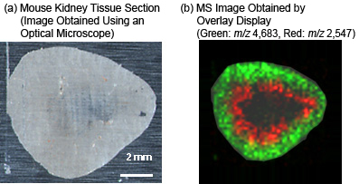
以下舉例說明胜肽及蛋白質的 MS 影像結果,這是來自大鼠腎臟組織切片 (冷凍切片),以 200 µm 間隔點樣基質 (芥子酸) 進行鑑定。
針對組織切片上 m/z 2,547 離子和 m/z 4,683 離子的 2D 分佈,以不同顏色標示,然後疊合生成 2D 分佈圖 (b)。
MALDI-TOF MS 影像能夠以視覺影像呈現組織切片特定質量的差異表現分子,例如胜肽及蛋白質。

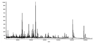
腎組織切片微區質譜圖

什麼是 MALDI-TOF MS 影像?
MALDI-TOF MS 造影影像採用一項 MALDI-TOF MS創新技術,可直接量測組織切片上的生物分子和代謝物,無需進行樣品萃取或標記。依據位置資訊及測得的訊號強度,可建立目標生物分子的 2D 分佈。使用 CHIP-1000 化學列印機進行預處理,以分配微量、皮升 (picoliter)、酶的濃度或 MALDI-TOF MS 基質,使 MALDI-TOF MS 造影達到絕佳的再現性及準確度。
Features of MALDI-TOF MS Imaging

- The CHIP-1000 Chemical Printer can dispense trace, picoliter, levels of a diverse range of reagents including matrix solutions and enzymes to facilitate MALDI-TOF MS imaging with excellent reproducibility.
- The CHIP-1000 enables MALDI-TOF MS imaging, identification, and structural analysis of diverse molecules, from lipids and low-molecular compounds to peptides and proteins.
- The acquired MS data can be read and analyzed using existing MS imaging software, such as BioMap (http://www.maldi-msi.org/).
- For details on MALDI-TOF MS, click here.


